Mda Mb 231 Cells

Cell micrograph cancer cell line mutation data transfex protocol mda mb 231 cells.
Mda mb 231 cells. Some cells in the culture usually. Transformed from human breast cancer cell line derived from metastatic breast cancer mammary gland epithelial cells mda mb 231 adhesive cell using lentivirus expressing firefly luciferase gene. The cell line demonstrates high level of bioluminescence signal via. In microarray profiling the mda mb 231 cell genome clusters with the basal subtype of breast cancer.
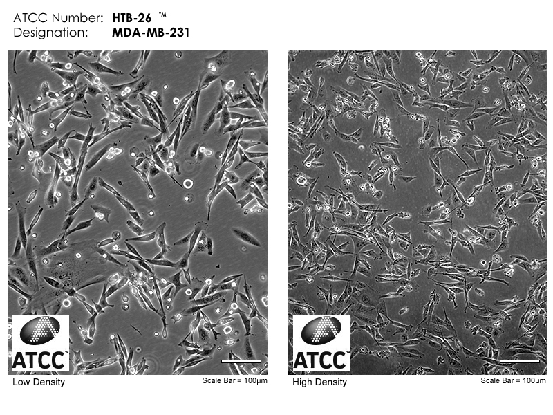
Faq s morphology of atcc htb 26 the atcc htb 26 cell line is epithelial like. Mda mb 231 cells showed both a stimulation of adenylyl cyclase and a plc dependent increase in intracellular ca 2 in response to nonselective adenosine receptor agonists. Mda mb 231 cells are grown at 37 c in leibovitz s l 15 medium supplemented with 2mm glutamine and 15 foetal bovine serum fbs. Scientists study the behaviour of isolated cells grown in the laboratory for insights into how cells function in the body in health and disease.
The luciferase was expressed under the enhanced cmv promoter. Experiments using cell culture are used for developing new diagnostic tests and new treatments for diseases. This cell line is er pr and e cadherin negative and expresses mutated p53. The puromycin marker was expressed under rsv promoter.
Brightfield images of cell aggregates and spheroids on day 4 were captured using evos m5000 microscope under 4x magnification. And subcultured when 70 80 confluent. The changes in the morphological pattern and gene expression of cells when grown in the presence of sera positive for anti carbonic anhydrase i ca i autoantibodies the effects of normothermic conditioned microwave irradiation on cultured cells using an irradiation system. Mda mb 231 has been used to study.
1 000 10 000 mda mb 231 cells were seeded for spheroid generation with or without collagen according to the above mentioned protocol. This is a list of major breast cancer cell lines that are primarily used in breast cancer. Dium supports the growth of this me cells in environments without co 2 equilibration. 3 both adenosine mediated responses in mda mb 231 cells were observed with the nonselective agonists 5 n ethylcarboxamidoadenosine neca and 2 3 hydroxy 3 phenyl propyn 1 yladenosine 5 n ethyluronamide phpneca but no.
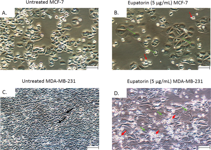
The cells are somewhat spindle shaped long and thin especially at subconfluence. Scale bar denotes 400 μm. Mcf7 cells are not metastatic and can serve as a negative.
